COME ABBIAMO IDEATO L'APPLICAZIONE
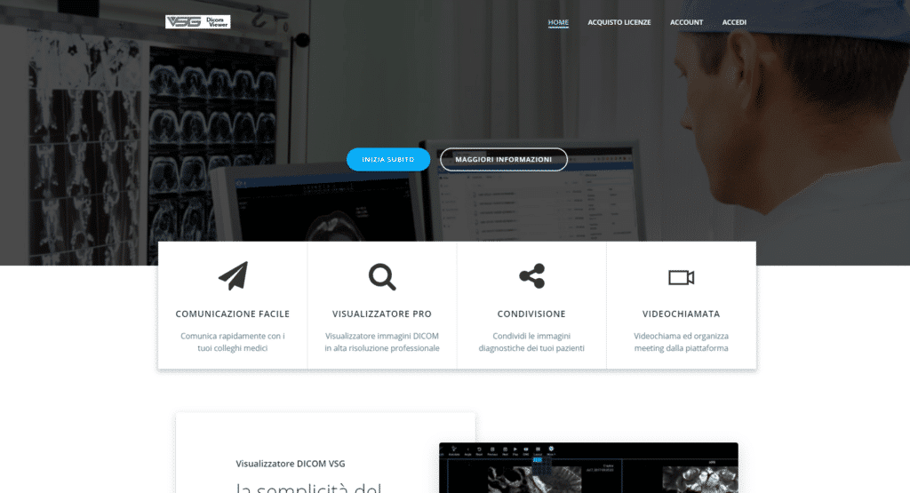
La nostra applicazione per la condivisione di diagnosi ed immagini mediche derivanti da Radiografie, Tac, Tomografie, ecc. e da qualsiasi tipo di macchinario medico che supporti il protocollo per immagini DICOM, è stata strutturata in tre sezioni principali:
- Sezione account, registrazione ed abbonamento
- Sezione gestione file e documentazioni
- Sezione Diagnosi e condivisione
In sostanza, il flusso è molto semplice e lineare seguendo questo schema: il dottore o il paziente si registra alla piattaforma tramite un abbonamento, che può essere settimanale, mensile, annuale o addiruttura per singola prestazione.
L’amministratore della piattaforma ovviamente decide che tipologia di abbonamento impostare e quali sono ovviamente i costi ed altri parametri, come i permessi relativi alla piattaforma.
L’abbonamento dà quindi accesso alla sezione account, dove il dottore o il paziente può controllare i suoi pagamenti, gli abbonamenti attivi, i giorni rimanenti alla scadenza, i suoi dati aziendali / del profilo e quindi controllare pienamente il suo account ed i suoi livelli di permesso.
Successivamente, il dottore o il cliente che utilizza la piattaforma può accedere al pannello di gestione files, dove potrà effettuare moltissime operazioni:
- Caricare i propri file medici online e renderli disponibili in qualsiasi momento (file DICOM)
- Caricare oltre ai file medici anche altri tipi di file (il sistema supporta quasi tutti i tipi di file, Word, Excel, PDF, immagini, video ed altro)
- Condividere tali file direttamente con altri membri della piattaforma, o invitare membri esterni alla piattaforma per condivisione mediante link protetti (da password) o non protetti.
- Fissare appuntamenti per le diagnosi tramite l’apposito calendario.
- Utilizzare lo strumento di MEETING, VIDEOCHIAMATA E CONDIVISIONE SCHERMO per condividere con altri utenti le proprie informazioni
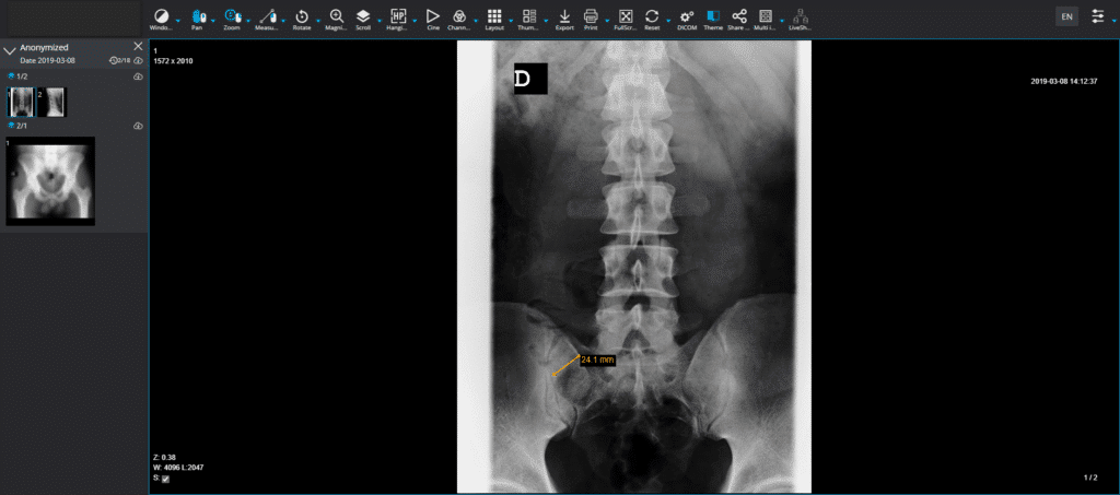
- Accedere al lettore medico DICOM per immagini radiografiche
Sono inoltre previste anche ulteriori funzionalità che verranno rilasciate eventualmente in versioni future della piattaforma.
L’ultima sezione di diagnosi molto tecnica utilizza il lettore per immagini DICOM ANCHE IN 3D utilizzato nelle macchine radiografiche, in terapia intensiva, ecc. ecc. ed ha la facoltà di leggere tali tipo di immagini ed effettuare misurazioni precise di quanto esaminato, come ad esempio nel caso di una ecografia. Può effettuare anche la renderizzazione e la visualizzazione di immagini radiografiche e tomografiche in 3D direttamente sullo schermo.
I dati del paziente possono essere poi condivisi in tempo reale con altri dottori, eliminando quindi i tempi di attesa che a volte possono essere molto lunghi.
L’applicazione è compatibile mobile e funziona come una web application, disponibile all’ indirizzo www.secondaopinione.net


